
If your teeth feel completely normal, do you really need dental X-rays? It is a reasonable question, especially because much of a dental examination appears to involve looking directly at the teeth and gums. However, some dental problems begin in areas that cannot be seen during a visual examination.
Dental imaging is one part of comprehensive preventive dentistry at Smart Dental in Richland. X-rays can provide information about areas between teeth, below existing restorations, around tooth roots, and within the supporting jawbone. That additional view can help your dentist identify changes that may otherwise remain hidden until symptoms develop.
Importantly, dental X-rays are not necessarily taken on exactly the same schedule for every person. The type and frequency of imaging should be based on your oral health, age, history, symptoms, previous dental work, and current risk factors.
What Can Dental X-Rays Show That a Visual Exam Cannot?
During a clinical examination, a dentist can see the exposed surfaces of your teeth, much of the gum tissue, existing restorations, and many other features of your mouth. However, teeth contact one another tightly, and their roots are surrounded by gum and bone.
Dental X-rays provide another layer of information. Depending on the type of image, they may help your dentist evaluate:
- Decay between neighboring teeth
- Changes beneath or around existing fillings and crowns
- The roots of teeth
- Bone levels surrounding teeth
- Areas of infection near a tooth root
- Developing or unerupted teeth
- Wisdom tooth position
- Certain jaw abnormalities
- Bone available for dental implant planning
Dental imaging does not replace a hands-on examination. Instead, the two provide different pieces of information that can be considered together.
Why Cavities Between Teeth Are Easy to Miss Without Imaging
Some cavities begin on chewing surfaces where they may eventually become visible. Others begin between teeth, where the surfaces contact and are difficult to inspect directly.
This is one reason bitewing X-rays are commonly used in preventive dentistry. They provide a view of the crowns of upper and lower teeth in the same area and can help reveal changes between teeth.
Finding decay while it is relatively small can matter because the size of a cavity influences how much tooth structure must be repaired. If damage remains localized, a tooth-colored filling may be sufficient. If decay grows significantly, the tooth may require a larger restoration.
Smart Dental also provides restorative dentistry in Richland when teeth need repair due to cavities, fractures, or other structural concerns.
What Are the Main Types of Dental X-Rays?
Different dental images answer different questions. Your dentist chooses the image based on what needs to be evaluated.
Bitewing X-Rays
Bitewings are commonly used to evaluate the areas between back teeth and to look at the level of supporting bone visible in that region. They are especially useful for detecting decay that may not be visible during a routine examination.
Periapical X-Rays
A periapical image generally shows the entire tooth, including the crown, root, and surrounding bone. It can be useful when evaluating tooth pain, possible infection, root conditions, or a specific area of concern.
Panoramic X-Rays
A panoramic image provides a broad view of the upper and lower jaws in a single image. It may be useful for evaluating developing teeth, wisdom teeth, overall jaw structures, and other larger areas that cannot be captured in one small intraoral X-ray.
Three-Dimensional Imaging
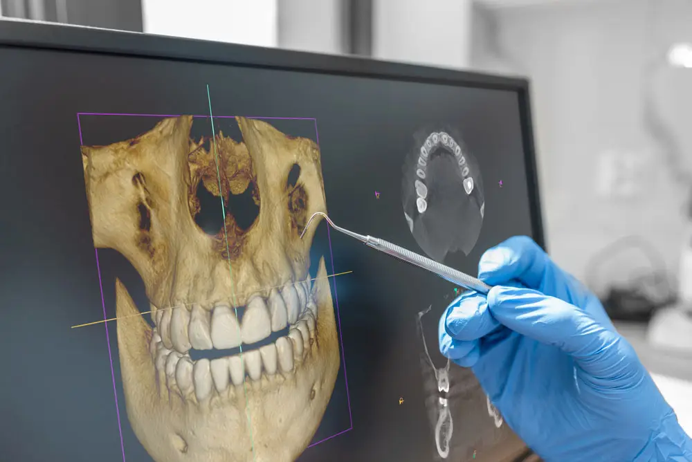
Three-dimensional cone beam imaging provides detailed views of dental and facial structures from multiple angles. It is not necessary for every routine dental examination. However, it can be valuable for procedures that require detailed anatomical planning, including certain dental implant and surgical cases.
For example, dental implant planning may require information about bone dimensions and the position of nearby anatomical structures that conventional two-dimensional images cannot show as completely.
How Often Should Dental X-Rays Be Taken?
There is no single schedule that is ideal for every patient. Someone with a history of frequent cavities may need imaging at different intervals than an adult with consistently healthy teeth and a low risk of decay.
Your dentist considers several factors when determining when new X-rays are appropriate, including:
- Your cavity history
- Current signs of tooth decay
- Gum and bone health
- Your age and stage of dental development
- Existing crowns, bridges, fillings, or implants
- Recent dental symptoms
- Previous images and whether meaningful changes are being monitored
- The type of treatment being planned
Therefore, asking why a particular X-ray is recommended is completely appropriate. Your dental team should be able to explain what information the image is intended to provide.
Do New Patients Always Need New Dental X-Rays?
Not necessarily. If you recently had dental X-rays at another office, those records may sometimes be useful to your new dentist.
However, whether older images provide enough information depends on their date, quality, type, and what has changed since they were taken. A new patient who has developed symptoms since the previous images may need updated or additional views.
If you are transferring dental offices, ask your previous provider about sending your recent records. Providing existing images can give your new dentist useful history and may prevent unnecessary duplication when those images are still diagnostically appropriate.
Are Digital Dental X-Rays Different From Traditional Film?
Digital dental imaging captures X-ray information electronically instead of relying on traditional film processing. Images can appear quickly on a computer screen, where the dental team can enlarge or adjust the display to examine specific areas.
This can also make patient education easier. Instead of simply being told that an area needs attention, patients may be able to look at the image while the dentist explains what is being monitored or treated.
Technology itself does not replace clinical judgment. Its value comes from combining clear imaging with an examination and a thoughtful treatment plan.
Benefits of Dental X-Rays
- Earlier detection: Imaging can reveal changes in locations that are difficult or impossible to see directly.
- Better treatment planning: Dentists can evaluate roots, bone, neighboring teeth, and other structures before recommending certain procedures.
- Monitoring over time: Comparing images can help identify changes in teeth or supporting bone.
- Evaluation of tooth pain: X-rays can provide information about structures beneath the gums when a tooth becomes sensitive or painful.
- Support for preventive care: Hidden decay may be identified before it produces obvious symptoms.
- Implant and surgical planning: Appropriate imaging can provide important anatomical information before procedures.
- Patient education: Digital images can help patients understand why an area is being watched or treated.
Can Dental X-Rays Find Every Dental Problem?
No single diagnostic tool can identify every oral health condition. Some cavities are easier to detect clinically, while certain cracks may not appear clearly on routine X-rays. Gum inflammation, bite problems, soft tissue changes, and many other concerns require direct examination.
Likewise, an unusual area on an X-ray may require additional testing before the dentist can determine exactly what it represents.
This is why good dentistry relies on several sources of information, including your symptoms, dental and medical history, clinical examination, gum measurements when appropriate, imaging, and sometimes additional diagnostic tests.
Why X-Rays Matter Even When Nothing Hurts
Pain is a useful warning signal, but it is not an ideal screening tool. A cavity can develop before it hurts. Bone changes can occur without obvious symptoms. An infection can sometimes remain quiet until it becomes more advanced.
Preventive dentistry is designed to identify concerns before they interfere with daily life whenever possible. Appropriate X-rays support that goal by giving the dentist information that cannot be obtained by looking at the tooth surface alone.
However, that does not mean more imaging is always better. The goal is appropriate imaging based on a clinical reason and your individual risk.
What If You Are Pregnant or Think You May Be Pregnant?
Always tell your dental team if you are pregnant or may be pregnant. That information helps the dentist consider the timing and necessity of dental imaging alongside your current dental needs.
Urgent dental problems should not simply be ignored during pregnancy. A painful or infected tooth may require diagnosis and treatment. Your dentist can determine which examinations and images are appropriate for the situation and coordinate care when needed.
Can X-Rays Help With Dental Emergencies?
Yes, dental imaging is frequently useful when a patient has unexpected tooth pain, swelling, trauma, or a broken tooth.
An emergency examination may require an image of the specific tooth or surrounding area to determine what is happening below the surface. For instance, pain can come from decay, infection, a damaged restoration, trauma, or other causes that require different treatment.
Smart Dental offers emergency dentistry in Richland for patients who need timely evaluation of sudden dental concerns.
Questions Patients Ask About Dental X-Rays
Why do I need X-rays if I have no cavities?
Your cavity history is only one factor. Dental X-rays can also provide information about tooth roots, supporting bone, existing restorations, developing teeth, and other structures that are not fully visible during an examination.
Can my dentist use X-rays from another office?
Often, yes, if the images are recent, clear, and show the areas your dentist needs to evaluate. Additional images may still be recommended when existing records do not answer the current diagnostic question.
Does every cleaning appointment include X-rays?
No. Imaging frequency should be based on individual need rather than automatically taking the same X-rays at every cleaning visit.
Why might I need a 3D dental scan?
Three-dimensional imaging may be recommended when detailed information about bone and nearby anatomical structures is needed for treatment planning, such as certain dental implant or surgical procedures.
Use the Right Information to Protect Your Smile
Dental X-rays are most valuable when they answer a specific question, reveal information that cannot be seen directly, or help your dentist monitor an area over time. Combined with regular examinations and professional cleanings, appropriate imaging can make preventive care more complete and treatment planning more precise.
If it has been a while since your dental exam, Smart Dental at 3200 Duportail St Suite 105 in Richland, WA can help you understand which preventive services are appropriate for your smile. Call Us Today at (509) 492-2051 to Schedule a Consultation, or Contact Us to request an appointment.